What is Cytology?
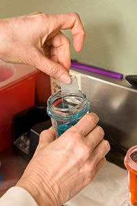
Staining Cytology Slide
What is cytology and how does it benefit the diagnostic process?
Cytology is simply the study of cells and is an extremely useful diagnostic tool that is used on a daily basis in veterinary dermatology.
Cytological samples are stained with special chemical stains to allow visualisation of the cellular and other material. We do this at our own on-site laboratory and can have the diagnosis with you in a matter of minutes.
Below, we’ve listed examples of situations where cytology is particularly useful:
Examination of discharges from the ear
This allows identification of organisms such as bacteria and yeasts as well as inflammatory cells. If rod shaped bacteria is seen through cytology, this is followed up by swabs being taken for bacteriology and antibiotic sensitivity testing.
Examination of surface skin cytology using acetate tape strip impressions
This is used primarily for the identification of yeasts often present in abnormal numbers in allergic skin disease.
Examination of pustule contents
Not all pustules are caused by infection. Auto-immune diseases such as pemphigus can present as a pustular disorder. Cytology of a pustule can reveal bacteria and white blood cells in a case of pyoderma, whereas a pustule in pemphigus will only show white blood cells and rounded epithelial cells, called acanthocytes.
Fine needle aspirate (FNA)
This is a method where cells are taken from masses such as tumours or those caused by infection with bacteria or fungal organisms. A sterile hypodermic needle is placed in the mass. Cells are then drawn in to the needle by applying a vacuum with a 10ml syringe.
The content of the needle is then sprayed on to a microscope slide, stained and examined closely. The common reason for using this technique is skin tumours can look like many other types of skin tumour. This technique is very useful in distinguishing between skin tumour types.
Impression smear
This simple procedure involves pressing a slide against an ulcerated skin lesion to pick up some of the cells on the surface that may help in making a diagnosis.

Pseudomonas rod shaped bacteria responsible for severe purulent otitis

Fine Needle Aspirate Histiocytoma

David Bentley at Microscope

Malassezia yeast organisms responsible for some cases of otitis and dermatitis
If you’ve noticed skin or ear problems in your pet, it’s vital that you contact specialised veterinary professionals before symptoms worsen. To book an appointment with our small animal skin and ear experts, simply call us below or email us on dgb@dermvet.co.uk.
