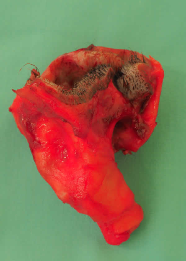
Total Ear Canal Ablation and Lateral Bulla Osteotomy (TECA-LBO) Surgery
It is a sad fact that not all cases of otitis can be dealt with medically. Otitis can be a very painful and debilitating condition and if it cannot be resolved by treating and reversing the primary and perpetuating factors, as veterinary surgeons we should offer to do whatever is needed to end the animal’s discomfort.
Removal all of all the diseased and painful tissues surgically is a very successful way of ending an animal’ s pain and discomfort in these situations. This is achieved by performing a Total Ear Canal Ablation and Bulla Osteotomy (TECA-LBO). The indications for a TECA-LBO are:
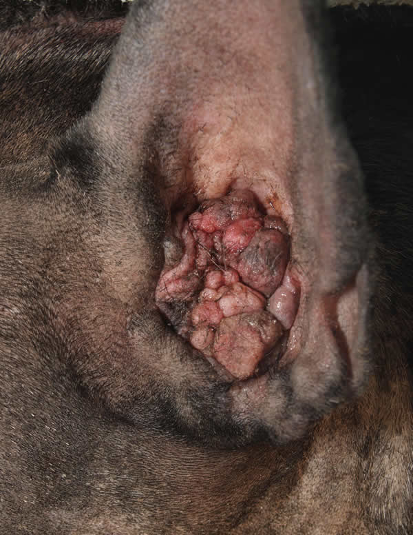
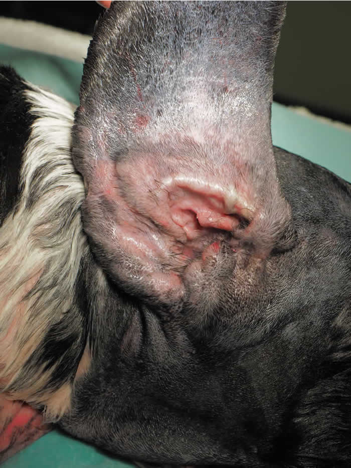
Chronic End Stage Otitis
End Stage Otitis ( where there is chronic irreversible change to the ear canal which prevents resolution of the pain, discomfort, infection)
- Intractable Otitis (where although the bulk of the ear canal is not “end stage” pathological changes in the ears of the middle ear, tympanic membranes and lower ear canal prevent complete resolution of the infection and healing of all affected tissues).
- Tumours in the ear canal which cannot be dealt with by lateral wall resection or vertical canal ablation.
- For financial reasons, sometimes the cost of this surgery can be less than a prolonged course of medical therapy, which can include several general anaesthetics and video otoscopy procedures, with no guarantee of success at the outset.
I have performed over 385 Total Ear Canal Ablations since 1991 and one thing that strikes me is how it can really transform dog’s life. Some of the common comments I get back from owners when their dogs come back for suture removal after this surgery include ” my dog is like a puppy again” or “I’ve got my dog back”.
The persistent pain and discomfort of living with a diseased ear canal just brings them really down, similar to a dog living with a bad mouth full of diseased teeth and gums where owners report similar radical changes in their dog’s demeanour after having a dentistry procedure/extractions.
TECA-LBO surgery is not a routine operation and there are risks attached to the procedure.
This is why you should always choose a surgeon who has great experience in doing the procedure and knows exactly what their complication rate is, or you should be referred to a surgical specialist at a specialist referral centre.
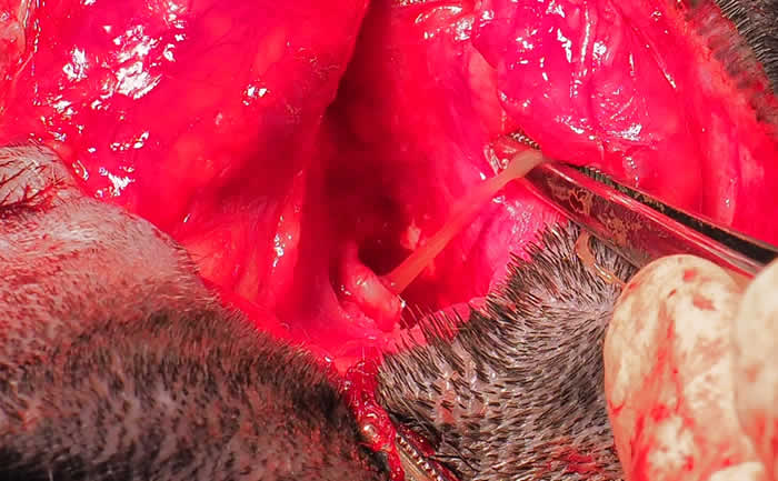
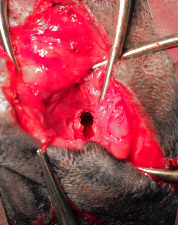
The surgery involves removing the whole of the vertical and horizontal canal, the integument that is adherent to the bone at the opening of the middle ear, the ear drum and part of the lateral wall of the middle ear (the “bulla osteotomy” part of the procedure), followed by thoroughly cleaning out the middle ear and removing the diseased middle ear lining with a curette.
The two main long-term complications were are concerned about are:
- Facial Nerve Paralysis
- Draining Tracts
Facial Nerve Paralysis The facial nerve, which supplies the muscles of the face and eyelids, comes out of the skull within millimetres of the junction of the ear canal and boney middle ear. This nerve has to be avoided during surgery because if it is cut , paralysis of those muscle will be permanent and there will be an inability to blink on that side and the lips will be flaccid on that side causing a facial droop and eating and drinking can be messy.
Fortunately most dogs mange to cope well with this. Sometimes false tear drops, are necessary to keep the affected eye moist but most dogs still manage to wipe over their eye when attempting to blink, due to movement of the 3rd eyelid when the eyeball retracts slightly during the blink process (due to different nerve).
Nevertheless, we don’t like to see this complication arising after surgery as it can look unsightly and the affected animals can remain as messy eaters. Temporary facial nerve paralysis lasting anything from a few hours to a few weeks can occur due to stretching of the nerve when retractors are used during the surgery to allow a good view of the surgical site.
Draining Tracts This is a much more serious complication which can occur as long as 2.5 years after surgery. It is vital that all integument, particularly the ear canal skin that is bonded onto the the bone for several millimeters into the opening of the middle ear (see diagram) is removed and all debris is removed from inside the middle ear along with the lining of the middle ear. Failure to do this can result in the development of a low grade infection deep underneath the surgical wound that can take along time to eventually come to the surface as a draining tract.
During this process painful thickening of the subcutaneous tissue can often be felt and the animal may experience pain when opening the jaw fully, so may be reluctant to eat or chew. If this occurs, surgical exploration of the site and re-curattage of the middle ear is usually necessary, but as the landmarks are not as obvious, there is greater chance of damaging the facial nerve.
Short Term Complications:
- Temporary Facial Nerve Paralysis
- Wound infection
- Wound breakdown (dehiscence)
- Vestibular dysfunction (inner ear problems leading to dizziness, inco-ordination, vomiting etc)
These are not common and are usually mild and last for only a few days (or weeks in the case of temporary facial nerve paralysis).
Clinical Audit
I have recently carried out a clinical audit on my complication rates for this procedure and compared it to what is published in the scientific literature. This was comparing my rate of complication with that performed by board certified surgical specialists at vet schools or referral centres. This was an audit of 55 operations between Nov 2011 ( when we changed our computer system) and Dec 2017. The results were as follows:
Summary of Post –Operative Complications
Total number of TECAs performed = 55
Complications Within First 12 Months of Surgery
1. Post-operative infection 11% (all of them mild)
2. Wound dehiscence 2%
3. Mild pinnal necrosis 2%
4. Transient facial nerve paralysis 9 %
5. Vestibular Symptoms 3.64 %
Complications Occuring over 12 Months of Surgery
1. Permanent Facial Nerve Paralysis 0%
2. Draining tracts/swelling requiring re-curettage of bulla 3.6%
3. Vestibular signs 0% These compare very favourably with that published in the literature, the zero rate of permanent facial nerve paralysis being excellent and the 3.6% rate of draining tract formation (2 cases) is at the lower end of the range that is published.
The two draining tracts cases resolved with a repeat surgery and have had no recurrenece with a 5 year follow-up. All the short-term complications were mild, only one requiring a minor surgical procedure to remove a small portion of necrotic pinna ( due to compromised blood supply in that area post surgery).
Click here for more info on my complication rate for Total Ear Canal Ablation Surgery
The cost of a TECA-LBO is between £2200-3000 per ear, including post-operative pain relief and antibiotics.
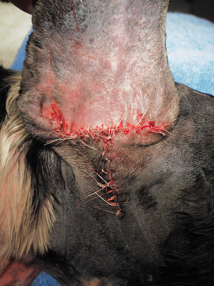
Recovery after TECA surgery is surprisingly fast. Most dogs are over the worst after 2 days of surgery and due to modern pain relief drugs don’t suffer too much discomfort in those first few days in any case. By the time they return for suture removal 2 week’s later their lives have been transformed.
Please ring our surgery on 0116 326 0402 or email enquiries@dermvet.co.uk if you wish to discuss Total Ear Canal Ablation and Lateral Bulla Osteotomy for your pet.

