These growths are very common on dogs and are called the “great imitator” as they can look very much like other skin tumours and skin lumps. The have to be considered as a differential whenever a dog is presented with a skin mass. Luckily in the majority of cases they are fairly easy to diagnose on cytology of fine needle aspirates taken from the mass. It is important to make a diagnosis as they are potentially malignant and ideally an excision margin of 2cm should be made when excising them prior to sending them to a dermatohistopathologist for grading.
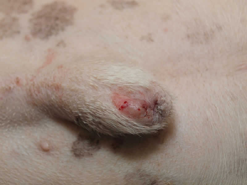
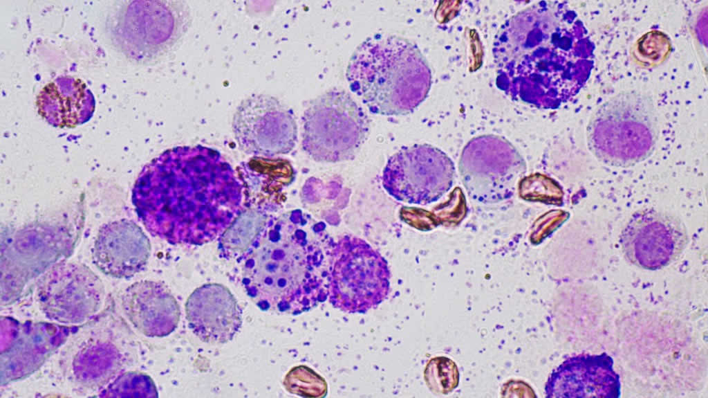
The growth on the prepuce of the dog in the left hand photo started as a small red spot and was thought initially to be either a bit of folliculitis or a mild injury. However, within 4 weeks the red spot had enlarged and trebled its size. A fine needle aspirate was taken from the lesion and examined microscopically after special staining. This revealed the typical mast cells containing dark staining granules inside the cells.
It was initially thought that the cytology indicated a grade 1, benign, MCT. Because of its site on the end of the prepuce, it was impossible to remove the lesion with a 2cm margin. The histopathology report came back with a diagnosis of a poorly differentiated MCT, possible malignant. Further radical surgery may be necessary , or if that is not possible, treatment with tyrosine kinase inhibitors such as mastinib (Masivet) or tocerinib (Palladia) can be useful at shrinking the tumours down.
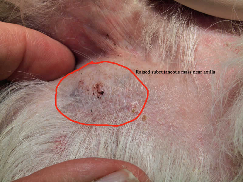
The photo on the right is from an 11 year old Peke. The soft, subdermal growth near the armpit almost had the appearance of a fatty tumour (lipoma) and had been slowly enlarging over 12 months , but fine needle aspirate suggested a poorly differentiated tumour with cells of various sizes and granularity extremely variable.
Cytology performed at the Dermvet Skin and Ear Clinic. From MCT on the right. Mast Cells of varying sizes, containing purple granules of varying sizes also
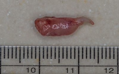
The growth was excised with a 2cm margin and surprisingly on the histopathology it was graded as well-differentiated and almost certainly benign!
The majority of canine mast cell tumours are benign, but the the ones that are malignant can spread to the lymph nodes and other organs. In these, thankfully, uncommon cases chemotherapy and radiotherapy can help to a degree but survival times are disappointing with most patients surviving approximately 12-18 months.
Inoperable tumours can sometimes be treated with the new Receptor Tyrosine Kinase inhibitors can sometimes produce dramatic results. It has been found that mast cells grow and proliferate is repsonse to specific cellular signals transduced by membrane bound receptors kinases such as c-kit. Mutations in c-kit signalling can results in the formation of a neoplastic population of mast cells. ReceptorTyrosine Kinase inhibitors such as toceranib (Palladia) and mastinib (Masivet) can inhibit c-kit mutants and can actually kill neoplastic mast cells. Results of treatment with these drugs are variable. Some dogs will see complete tumour regression others a reduction in size and some other no response at all.
In one clinical trial of toceranib* , the overall response rate of 145 dogs was 42.8% (21 complete responses and 41 partial responses), with an additional 16 dogs experiencing stable disease. Dogs with MCT that had KIT mutations were more likely to respond to toceranib than those without (69 versus 37%).

Nasopharyngeal Polyps in Cats
Ear disease isn't that common in cats. Apart from infestation with ear mites (otodectes) which in my practice is most commonly seen in newly purchased kittens, otits externa, which is often seen in dogs, is only seen occasionally. Very often , when I am referred a...
A Case of Primary Malassezia Dermatitis
We recently saw a rare case of Primary Malassezia (Yeast) Dermatitis in a young Dachshund
Demodicosis
I regularly see dogs suffering with various presentations of demodicosis, a skin condition caused by….

