One of the primary caused of otitis externa that we see in dogs and cats is a tumour in the ear canal. Tumours inside the ear cause otitis by occluding the ear canal and changing the microenvironment , creating favourable conditions for bacteria and yeasts to proliferate and eventually leading to infection, discharge and pain. In the past, the only way of dealing with these tumours was to either remove them with biopsy forceps via video otoscopy if they were very small or with major surgery, either a lateral wall resection, vertical canal ablation or sometimes a total ear canal ablation (TECA-LBO) if they were larger. Even if small tumours could be removed it was difficult to be sure that all tumour tissue had been removed and if the tumour was malignant then it could recur and still risk spreading to other parts of the body.
Another way of treating these tumours is to use a laser directed to the site via an optical fiber that can be passed down the channel of a video otoscope. The laser can then be used to vaporise the tumour in situ if it is small or to treat the remnants of the tumour after it has been debulked by traction or by the use of fexible biopsy forceps to cut it away from the ear canal wall.
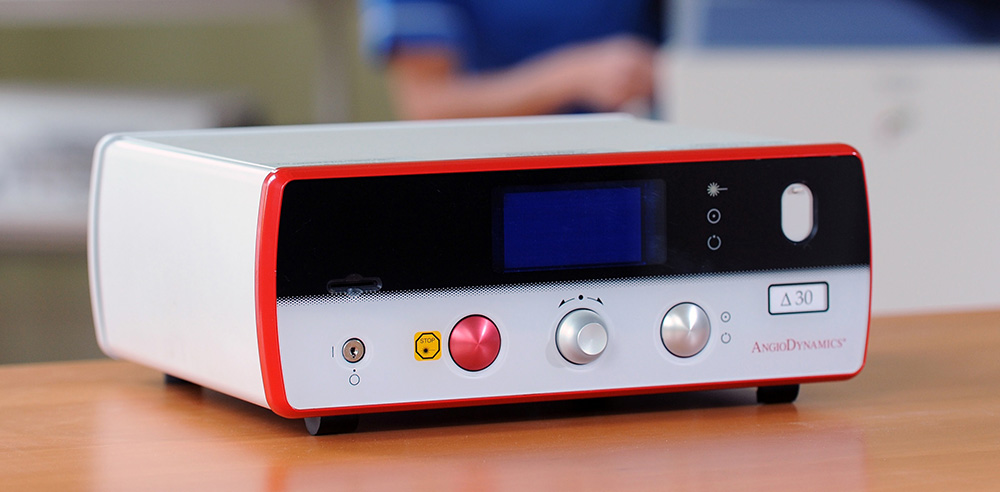
For the last 5 years I have been using a Diode Laser from UK company Excel Lasers has meant that I have been able to offer alternatives to much more expensive and risky sharp surgery for the removal of ear tumours and other lesions inside the ear canal. The laser energy is passed down a 600microm wide optical fiber from the unit pictured, to produce an extremely localised area of intense heat that will cut, coagulate or vaporise tissue with minimal damage to surrounding tissue.
Here is a video of a recent case I treated. Charley had a Ceruminous Gland Adenocarcinoma almost completely occluding the ear canal and there was a secondary pseudomonas otitis. After debulking the tumour, which luckily had a fairly narrow base, and waiting for the histopathology report, I went back in with the laser to destroy any remnants of this malignant tumour which would have been present.
This diode laser is also an excelent tool for treating other lesios in the ear canal such as:
- Cerumuminous gland adenomatosis in cats
- Polypoid ceruminous gland hyperplasia in dogs – this is where this laser will getting most of its use. It can speed up the resolution of this pathology which offten occurs in chronic otitis and although can sometimes be treated successfully with steroids over several weeks, does not always clear up. Laser ablation of theses lesions is a much faster and more effective way of dealing with theses lesions which are a perpetuating factor in otitis.
More videos will be posted showing the laser being used to treat these two conditions.

Feline Ceruminous Gland Adenomatosis

Polypoid Ceruminous Gland Hyperplasia
Laser Removal of Lesion Attached to Ruptured Ear Drum
Alice has a history of otitis of several months’ duration. Her ear drum was ruptured and there was a fleshy growth attached to the ruptured edge. The growth was removed with biopsy forceps and sent off for histopathology. The remnants of the growth were ablated using the diode laser. Healing was rapid and within 2 weeks the ear drum was completely healed. Histopathology showed this mass to be exuberant granulation tissue and not a tumour.
Polypoid Hyperplasia- Successful treatment with laser over several months- NO TECA-LBO NECESSARY
Patch came to see me with a very long history of otitis of over a year’s duration with a ruptured ear drum in one ear. This case would normally have been sent straight for surgery for a bilateral TECA-LBO , but his owners were keen to try avoid surgery if at all possible, It took 4 separate sessions with the laser via video otoscopy to successfully remove all hyperplastic tissue and get the otitis cured. He is now doing very well just using hydrocortisone aceponate spray in the ear canals twice weekly
Since October 20024 I have now added a Vetscalpel CO2 laser to my armoury, which will be used from now on for the majority of laser surgery cases inside the ear canal. However, the diode laser will still have its place in treating some of the smaller lesions at the very bottom of the ear canal, where it will be more accurate and safer to place the fibre optic tip on the lesion directly rather than using an invisible beam of laser light that may be too risky to use near the ear drum.

David Bentley and Kerry Kooi from Vetscalpel Inc. on the day I was handed over my new C02 laser
Here is one example of it being used for treating Feline Cystomatosis. As you will see, because the CO2 laser cannot be used with water (as the diode laser can) the video isn’t as crystal clear as the ones showing the diode laser in use, due to the smoke that is produced and risk of wax etc smearing on the front of the video otoscope lens. However, the two great advantages of the CO2 laser over the diode laser are that there is far less collateral thermal damage to the surrounding tissue and larger lesions can be ablated much faster than with the diode laser.