Cases of Demodicosis (Demodectic Mange)
I regularly see dogs suffering with various presentations of demodicosis, a skin condition caused by the canine species of demodex mites.
These mites are considered to be a normal part of the skin fauna, and it is thought that puppies acquire these mites from their mothers in the first few days of life. These mites live within the hair follicle or the sebaceous glands and they have 4 pairs of stumpy legs and a long slender body so they look more slug-like.
Although it is thought that all dogs carry these mites, it is only usually possible to find these mites on skin scrapings in individuals suffering with demodicosis, as a the mites are present in very high numbers in these cases. Demodex canis is the cause of the majority of cases in dogs, although cases are also seen with the longer bodied mite Demodex injai and the short bodied Demodex cornei ( may actually be a avriant of D.canis) can also be seen is some cases.


Juvenile-onset demodicosis can be either localised or generalised and the course of the disease and prognosis is vastly different. Certain breeds are predisposed due to genetic factors and these include Bull breed dogs, Chinese Sharpeis and Westies and Scottish Terriers.
The localised form presents as one or a few more small solitary slightly red patches of alopecia that are not itchy. these often occur on the face or forelegs. The majority of these cases self cure and do not need treating.
Occasionally these only occur in the ear canals causing a waxy otitis and theses cases will need treating. The generalised form presents as large area of the body affected, often including the limbs, feet and head.
The definition of localised versus generalised has been a matter of debate but recently, in the journal Veterinary Dermatology, a panel of experts led by Professor Ralph Mueller considered demodicosis to be localised if there are no more than four lesions with a diameter of up to 2.5cm (Treatment of Demodicosis in Dogs: 2011 Clinical Practice Guidelines.- Mueller et al Veterinary Dermatology 23 pages 86-96 July 2012 – Wiley-Blackwell)
 Demodicosis")


Left untreated this can be a fatal disease and in the past many owners elected to have their affected dogs put to sleep.
Some individuals have the demodicosis confined to the feet and these are often affected with a bacterial pyoderma as well. These can be difficult to treat.
Generalised demodicosis is also complicated by the fact that the follicles become infected with bacteria, usually Staphylococcus pseudintermedius and is some cases this can cause pustules to form on the skin, so called pustular demodicosis. The skin develops alopecia, crusting, thickening and hyperpigmentation, and because of the concurrent infection, the lymph nodes are often enlarged and the dog can be systemically ill. Left untreated this can be a fatal disease and in the past many owners elected to have their affected dogs put to sleep.
Some individuals have the demodicosis confined to the feet and these are often affected with a bacterial pyoderma as well. These can be difficult to treat.
Diagnosis
Diagnosis is made by finding mites on microscopic examination of deep skin scrapings. Very often, numerous mites in all stages of development, i.e. adults, nymphs and eggs can be seen on the one slide, but in some individuals, particularly dogs with very thick skin, only an occasional mite can be found. The mite can also be found attached to the roots of hairs when they are plucked and this method is often used here at the Dermvet Skin and Ear Clinic in areas where skin scrapings are difficult to perform e.g around the eyes and between the toes. Very occasionally, diagnosis can only be confirmed on skin biopsy.
As some cases can be missed due to low mite numbers it is always a good idea to seek a veterinary dermatologist if you have a dog with skin disease that is not responding to treatment.
Treatment
Juvenile-onset demodicosis will often spontaneously recover. Juvenile -onset generalised cases always need treating as spontaneous recovery only occurs in around 30% of cases less than a year of age and they are usually complicated by bacterial infection, so those cases would still need at least a course of antibiotics. Treatment sometimes has to be given for months and in a few cases, has to be continued for life as they relapse when treatment is terminated.
A search for an underlying cause has to be made in adult-onset cases as if this is not treated, relapse is likely. Causes include prolonged use of steroids for treatment of allergies or autoimmune disease (I have seen several cases due to this recently), Cushings disease, hypopthyroidism, malignancies and other disorders such as liver or renal failure.
Treatment involves using drugs that kill the mites and treating the associated pyoderma with antibiotics and antibacterial shampoos. Antibiotic therapy with bactericidal antibiotics can be necessary for as long as 6-8 weeks.
Isoxazolenes
In the last year or so (2016) there has been a revolution in how we can successfully treatment of canine demodicosis. Currently there are only two veterinary products licensed for the treatment of demodicosis , which are an Amitraz dip which is messy, smelly and requires weekly dips for up to 12 weeks, or a spot-on containing moxidectin weekly. The spot-on is not very effective at curing a case as although it lowers mite numbers is rarely results in 100% mite reduction.
In the last 2-3 years, new products of the isoxazolene group of drugs have come onto the veterinary market for the treatment of fleas and ticks. One of them, Fluralaner, (Bravecto) has been shown to be very effective, with 100% mite reduction after 56 days of one tablet. https://parasitesandvectors.biomedcentral.com/articles/10.1186/s13071-015-0775-8.
Despite this study and many others, confirming its efficacy against demodex, a product licence is not yet available, but will be in time. A related product, Sarolaner (Simparica) is curently licensed for treatment of tick, fleas and sarcoptic mange mites and shows equal efficacy to Bravecto. https://www.sciencedirect.com/science/article/pii/S0304401716300504
As theses are safe drugs, already licensed in dogs for the treatment of fleas, ticks and sarcoptic mange, we are now using these drugs in preference to the unlicensed treatments, such as daily oral ivermectin or milbemycin, which although very effective, took months to work in may cases and ivermectin , in particular, couldn’t be used in all dogs as some with a particular gene, were at risk of toxic effects which could be fatal.
I include these older treatments for reference, but is unlikely we will have to use them in future.
Ivermectin
This is a drug used in cattle, sheep and pigs to treat parasites. It will never be licensed in dogs as it is dangerous to use in dogs with the MDR1 gene ( found in some collies and other herding dogs and occasionally in other breeds).
If the drug is given to dogs with this gene they suffer neurological side effects that can be fatal. There are two ways of going about treating with this drug. One is to have the gene test, which costs around £80 or the other is to gradually work up the dose to the required level over a period of 7-10 days starting at 10% of the end dose and checking for any mild neurological signs (which would be reversible).
I have treated many non-collie type dogs with this protocol and have only had to stop treatment due to neurological signs in a couple of cases. In both cases, the signs were very mild and cleared once stopping treatment. The drug is given orally, daily for at least a month after multiple skin scrapings are negative. Signed owner consent is required if we use this drug.
Milbemycin
This is in the same family of drugs as Ivermectin and is used in several veterinary worming products. It as safe to use in dogs with the MDR1 gene. The drug is not freely available in the UK without being combined with other drugs such as praziquantel ( for tapeworms) or spinosad (for fleas).
It is available as a sole drug in Europe and America for the treatment of heart worm but a license has to be obtained from the Veterinary Medicines Directorate to import it, which adds to the expense of an already very expensive drug (10x the expense of Ivermectin). In the UK, it can be used off-license with informed signed consent in the form of the the cat worming tablets containing milbemycin and praziquantel.
The amount of praziquanatel relative to the milbeycin in the cat version of these tablets is much less that in the dog version and is safe when the correct dose of Milbemycin is given. As with Ivermectin, treatment has to continue for at least 4 weeks beyond multiple negative skin scrapings. Although safe, this is an expensive treatment that can cost over £300 a month for a 20kg dog.
If your dog’s skin is showing signs of alopecia, inflammation, scaling or if your dog is itching or scratching and you want to see a veterinary dermatologist in the east midlands or west midlands, contact us at the Dermvet Skin and Ear Clinic on 0116 326 0402 or email dgb@dermvet.co.uk


Nasopharyngeal Polyps in Cats
Ear disease isn't that common in cats. Apart from infestation with ear mites (otodectes) which in my practice is most commonly seen in newly purchased kittens, otits externa, which is often seen in dogs, is only seen occasionally. Very often , when I am referred a...
A Case of Primary Malassezia Dermatitis
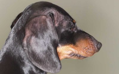
We recently saw a rare case of Primary Malassezia (Yeast) Dermatitis in a young Dachshund
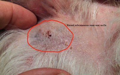
2 Cases of Mast Cell Tumours
These growths are very common on dogs and are called the “great imitator” as they can look very much like other skin ….