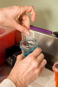
Diagnostic and Lab Testing
We complete thorough diagnostic tests to diagnose tricky conditions and offer your pet the best solutions and treatment
 Many skin and ear conditions can look very similar to each other resulting in difficult diagnosis. Veterinary dermatologists will not only have to use their knowledge of allergies and other conditions, but also complete a variety of diagnostic tests to get to the bottom of the problem.
The first thing that our dermatology specialists have to do is eliminate possible parasites, infections and allergies. Correct and safe treatment can’t be given without the correct diagnosis and this is particularly important when dealing with conditions that are incurable, such as atopic dermatitis.
We complete a range of tests on animals that come to us with possible ear and skin problems. Skin scrapings and cytology tests are performed on the majority of skin cases while other diagnostic and laboratory tests such as trichograms, skin biopsies, allergy tests and blood tests are performed from time to time.
Our state-of-the-art on-site laboratory means that we can offer your pet fantastic and thorough treatment. Without it, diagnosis and treatment of some conditions may take longer to complete meaning the symptoms could worsen. The laboratory enables to act quickly and test anything that we feel requires close attention.
It’s vital that we have complete evidence of a condition before we can offer suitable treatment. The wrong type of treatment can often mean that your pet needs to take inappropriate medication for the rest of their lives.
We’ve listed the common types of diagnostic and laboratory tests that we regularly complete below.
See the types of cases that these procedures may be used for:
What is a skin biopsy and how does it help the diagnostic process?
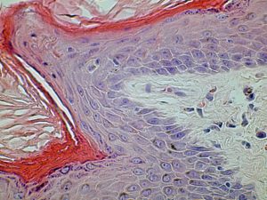
Histology Skin
A skin biopsy is a technique in which skin lesions are removed and examined closely using a microscope by a dermatohistopathologist. Although many skin conditions can look similar to the naked eye, under the microscope there can be many differences in appearance which allow us to offer a definitive diagnosis or at least rule out other conditions.
When we remove theses skin lesions, we send them away to a dermatohistopathologist for examination. There are only a very small number of these skin pathology specialists in the UK and we have a great working relationship with several of them.
Cases that often require skin biopsy include those that require investigation of hair loss, in disorders whether there is a lot of scale production, ulcerative skin diseases, suspected autoimmune skins diseases or when a tumour is removed.
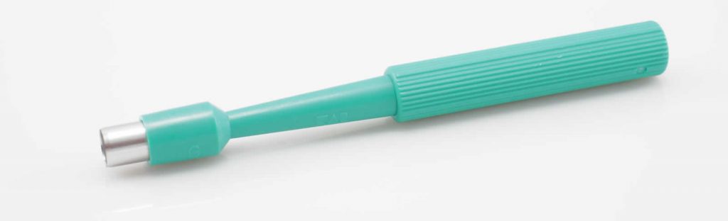
Biopsy Punch
Skin biopsies are performed either under general anaesthesia or under sedation.
Most cases will involve taking several punch biopsies, where a small piece of full thickness skin between 4-8mm in diameter is removed and placed in fixative solution.
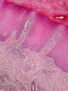
Histopathology – cat with footpad hyperkeratosis
The samples are then sent to the lab where the tissue is processed.
The biopsy is embedded in paraffin wax and sections one cell thick are sliced off and mounted on a microscope slide. They are then stained with special stains prior to being examined.
As soon as we receive the results of the biopsy, we’ll notify you and endeavour to explain the result in as simple terms as possible. We’ll also notify you of the next course of action for your pet.
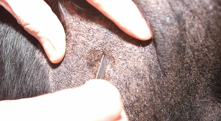
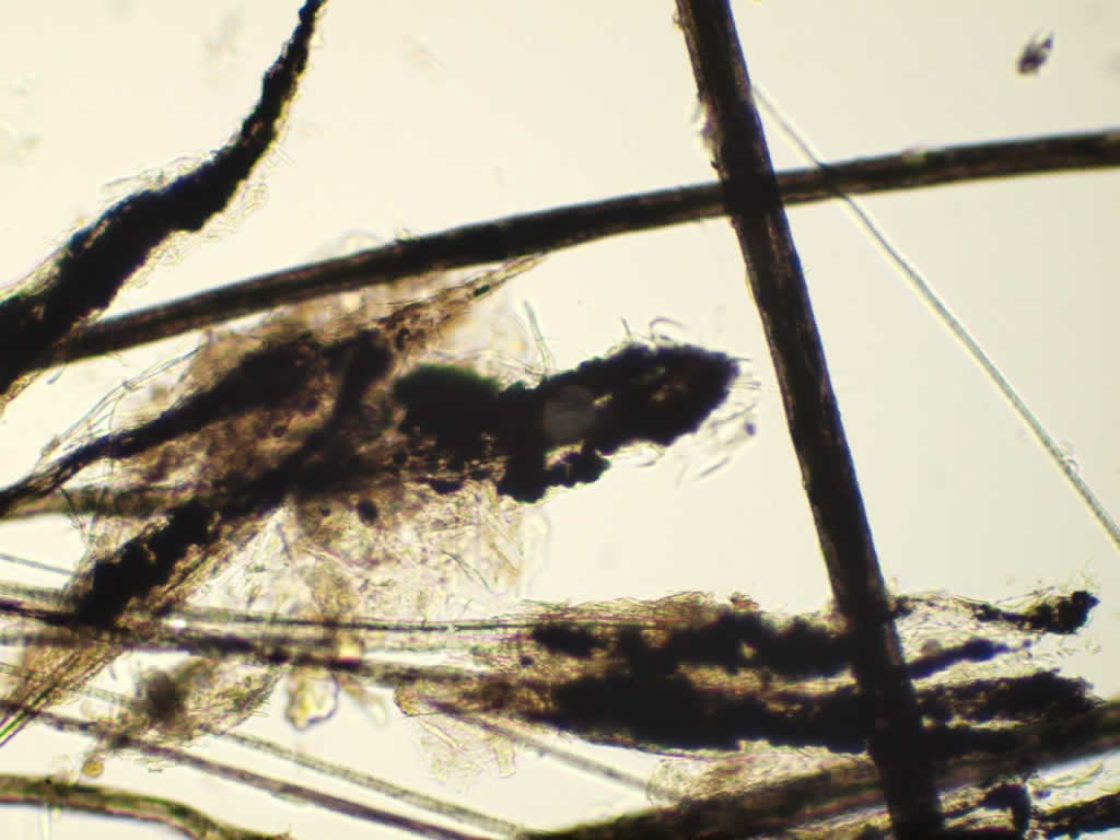
Why do we have to perform skin scrapings at the outset of a dermatological diagnostic work-up?
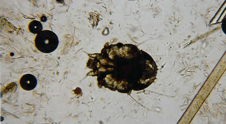
Skin scrapings are necessary for the majority of dermatological diagnostic work-ups. Diseases such as demodectic mange can usually easily be diagnosed by this method.
A small amount of liquid paraffin is placed onto the skin and the skin is then gently scraped until there is oozing of blood from capillaries. Scrapings have to be this deep as these demodex mites live deep down inside the hair follicles.
Mites can also sometimes be found from hair plucks if the roots are examined in liquid paraffin under the microscope. More superficial scrapings will be used to find the mites causing sarcoptic mange (scabies) or when there is Cheyletiella (Fur Mites). These mite infestations can often look like an allergy case as they can result in itching and scratching, so it is imperative that parasites are ruled out at the outset.
If these steps are missed or not performed properly it could result in incorrect treatment being given to your pet. We have seen several cases of atopic dermatitis that have been on steroids for a long time and have flared up after being under reasonable control. This is due to the development of adult-onset demodectic mange as a consequence of being on steroids.
Without performing skin scrapings and making the diagnosis it could have resulted in more steroids being given and making the condition even worse.
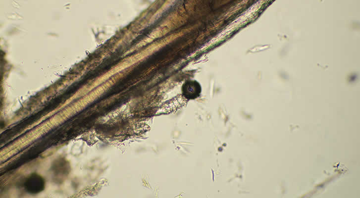
Demodex hair root
Demodex canis mite adjacent to hair root
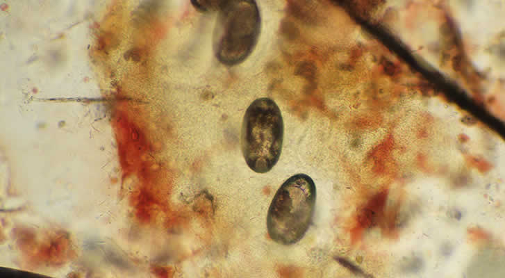
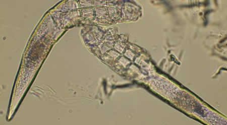
Sarcoptes scabiei eggs
Sarcoptes scabiei
Skin Scraping
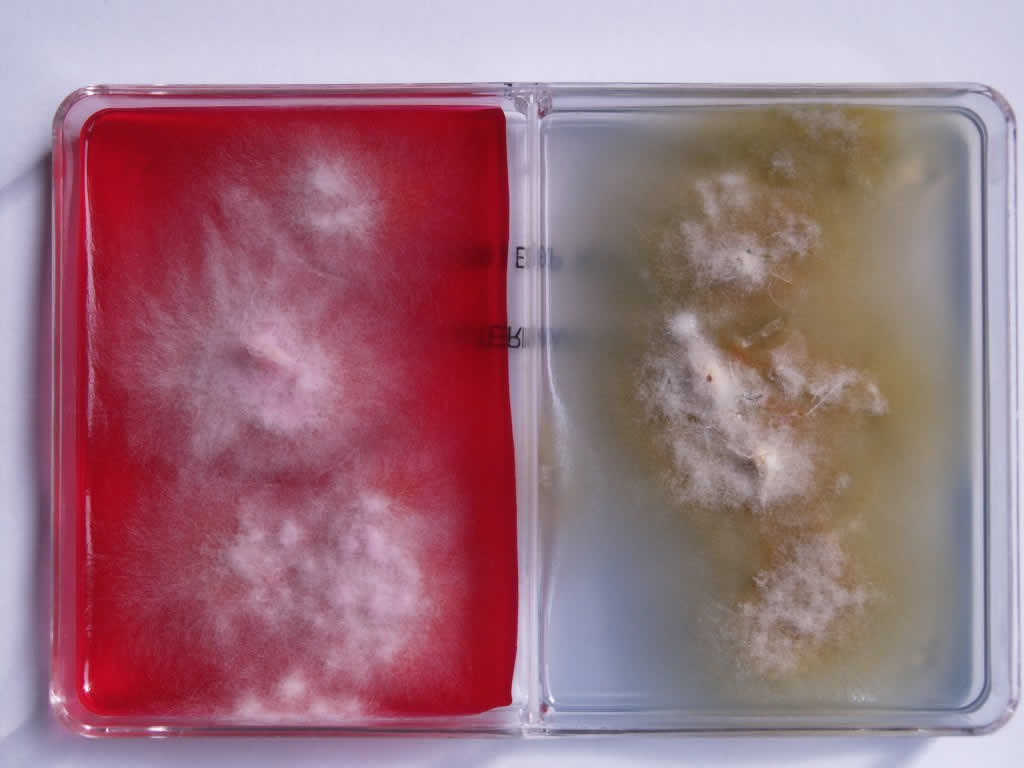
When do we have to perform a Fungal Culture?
Many cases of alopecia and scaling, both generalised and focal, include fungal infection with dermatophytes (ringworm) as one of the differential diagnoses. Fungal culture is often performed as part of a work of cases such as these, particularly in cats.
We use a special type of dual culture containing Dermatophyte Test Medium (DTM) and Enhanced Sporulation Agar (ESA). The DTM turns red in the presence of a dermatophyte and the ESA encourages the formation of special spores, called macroconidia.
This enables us to determine the species of dermatophyte causing the infection from its shape and number of cells when viewed under a microscope. It is surprising how many cases of ringworm in cats can look like other skin diseases, so many of them will have a fungal culture as part of their work-up.
Fungal Culture
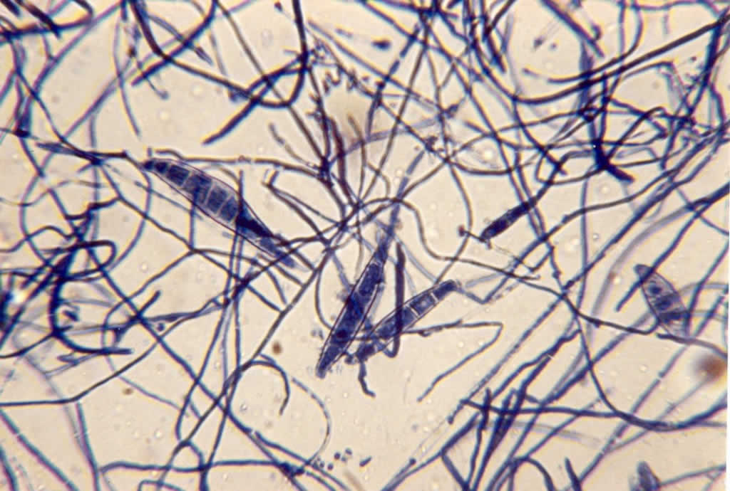
Macroconidia Microsporum Canis
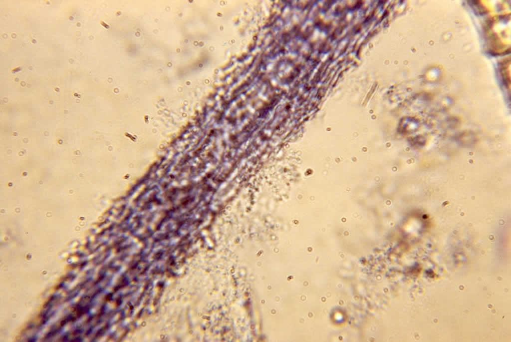
What are trichograms and what use are they in making a diagnosis?
Quite simply, a trichogram is the microscopic examination of hair shafts. Quite a lot of information can be gleaned from this simple test. For instance, if a cat is losing its fur and there is no inflammation we can often prove that the fur is being chewed or licked off, as the ends of the hair shafts are frayed or blunted rather than tapering to a point. Certain diseases, such as black hair follicular dysplasia and colour dilution alopecia, can be determined by the appearance of abnormal clumping of melanin (pigment) granules in the hair shafts and roots.
Causes of ringworm can be diagnosed sometime by finding the fungal filaments invading the hair shaft if special stains are used. Hormonal alopecia will have the hair roots all in the same stage of the hair cycle.
Ringworm
Black Hair Follicular Dysplasia-showing abnormal melanin clumping
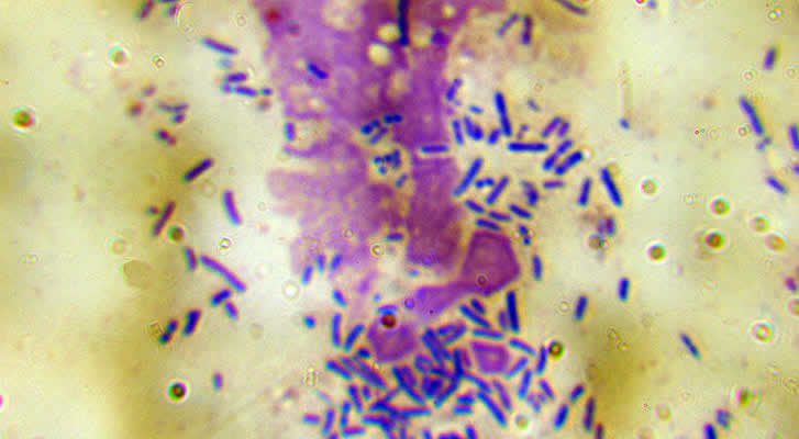
What is cytology and how does it benefit the diagnostic process?
Staining Cytology Slide
Cytology is simply the study of cells and is an extremely useful diagnostic tool that is used on a daily basis in veterinary dermatology.
Cytological samples are stained with special chemical stains to allow visualisation of the cellular and other material. We do this at our own on-site laboratory and can have the diagnosis with you in a matter of minutes.
Below, we’ve listed examples of situations where cytology is particularly useful:
Examination of discharges from the ear
This allows identification of organisms such as bacteria and yeasts as well as inflammatory cells. If rod shaped bacteria is seen through cytology, this is followed up by swabs being taken for bacteriology and antibiotic sensitivity testing.
Examination of surface skin cytology using acetate tape strip impressions
This is used primarily for the identification of yeasts often present in abnormal numbers in allergic skin disease.
Examination of pustule contents
Not all pustules are caused by infection. Auto-immune diseases such as pemphigus can present as a pustular disorder. Cytology of a pustule can reveal bacteria and white blood cells in a case of pyoderma, whereas a pustule in pemphigus will only show white blood cells and rounded epithelial cells, called acanthocytes.
Fine needle aspirate (FNA)
This is a method where cells are taken from masses such as tumours or those caused by infection with bacteria or fungal organisms. A sterile hypodermic needle is placed in the mass. Cells are then drawn in to the needle by applying a vacuum with a 10ml syringe.
The content of the needle is then sprayed on to a microscope slide, stained and examined closely. The common reason for using this technique is skin tumours can look like many other types of skin tumour. This technique is very useful in distinguishing between skin tumour types.
Impression smear
This simple procedure involves pressing a slide against an ulcerated skin lesion to pick up some of the cells on the surface that may help in making a diagnosis.
Pseudomonas rod shaped bacteria responsible for severe purulent otitis

Fine Needle Aspirate Histiocytoma
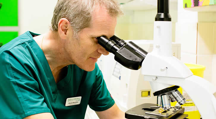
David Bentley at Microscope
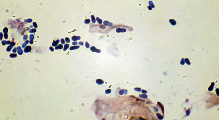
Malassezia yeast organisms responsible for some cases of otitis and dermatitis
Blood Tests
 Investigation into the causes of some skin conditions will involve taking blood samples for various tests. Some skin conditions are a marker for internal disease , for instance adult -onset demodicosis (demodectic mange) which can develop as a consequence of altered immune system function brought about by diseases such as Cushings Disease.
A full blood work up will involve checking a full blood biochemistry and haematology profile. Usually we can do this in-house and have the results within minutes. When hormonal diseases are suspected in cases such as alopecia or recurrent bacterial skin infections, we may take samples to be sent to an external laboratory for thyroid profiles and/or adrenal function tests, with resuklts usually available within 24 hours.
Blood collection is usually straightforward and most animals tolerate the sampling procedure very well.
Woods lamp examination
When checking animals for dermatophytosis (ringworm) we often will examine them under a special type of ultraviolet light called a Wood’s lamp. 50% of strains of Microsporum canis, the most common cause of ringworm in cats, will show the infected hairs fluoresce apple green under this lamp.
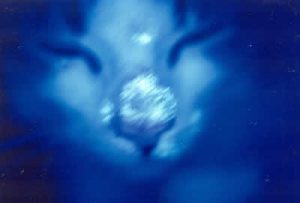 We still have to rely on fungal culture to confirm the diagnosis, but the lamp does have its place in checking animals which are known to be infected with a fluorescing strain and we can start treatment immediately if a case is positive as it may take up to 2-3 weeks to rule out a suspected case based on fungal culture.
Here is a rather blurred photo of a cat infected with Microsporum canis showing the bright apple green fluorescence of the infected hairs.
To function properly the lamp has to warm up over 5 minutes and examination has to take place in a darkened room.
If you’ve noticed skin or ear problems in your pet, it’s vital that you contact specialised veterinary professionals before symptoms worsen. To book an appointment with our small animal skin and ear experts, simply call us below
